Domanda n. 11
Carmen Gonzales, MD and Lois Hart, RDMS
Massachusetts Eye and Ear Infirmary, Harvard Medical School, Boston, MA
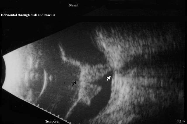
Fig. 1
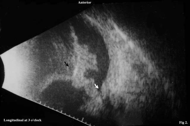
Fig. 2
Figg. 1-2: Ultrasound of a 67 year old patient complained of gradual loss of vision OS over one week peroid of time. History of DM for three years. No previous ocular surgeries. On exam, there is no view of the fundus due to vitreous hemorrhage. Ultrasound is requested to rule out posterior segement pathology.
- Describe the findings of the B-scan ultrasound
- What would your first diagnostic interpretation of this exam be and what
is the correct diagnosis?
- What ultrasound features of the exam strongly suggest the diagnosis?
- What clinical information would be important in your diagnosis?
- What would be the best clinical approach?
RISPOSTE
- Describe the findings of the B-scan ultrasound
Answer: The scan reveals a phakic globe within normal contour. Thickened, irregularly contoured, moderate to high reflective, V-shaped membrane which appears as a closed funnel posteriorly with a point of attachment at nasal peripapillary region. Low reflective echoes noted both anterior and posterior to this membrane.
- What would your first diagnostic interpretation of this exam be and
what is the correct diagnosis?
Answer: The first impression is that of a total funnel retinal detachment which is closed posteriorly with subretinal debris. The correct answer is posterior vitreous detachment (PVD). Hemorrhage in the vitreous cavity may deposit around the PVD giving the thickened and irregular appearance of this membrane.
- What ultrasound features of the exam strongly suggest the diagnosis?
Answer: The insertion site of the membrane into the nasal peripapillary region and not directly on the disc suggests the PVD. Kinetic information may also be helpful.
- What clinical information would be important in your diagnosis?
Answer: Patient with retinal detachments often complain of sudden visual disturbances described as "curtains" and presence of photopsias. Myopia and trauma are other risk factors for retinal detachments. The diabetic history in this patient may be misleading since vitreous hemorrhage with proliferative vitreoretinopathy and tractional retinal detachment may be expected. If the patient has had many previous ocular surgeries, the usual anatomical distribution of membranes within the eye may have changed.
- What would be the best clinical approach?
Answer: This patient was observed for a few months while photocoagulation was given to treat a proliferative diabetic retinopathy in the fellow eye. As spontaneous reabsorption of the vitreous hemorrhage did not occur, vitrectomy was performed.
- Describe the findings of the B-scan ultrasound