Domanda n. 3
a cura di:
Rosa Y Kim, MD
Massachusetts Eye and Ear Infirmary, Harvard Medical School, Boston, MA
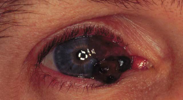
Figure 1: This is a 43 year old HIV+ man with a growth on the right eye for 4 months.
- What is the differential diagnosis of this lesion?
- When one observes pigmented lesions of the conjunctiva, what are some useful historical questions that should be asked?
- What pre-existing conditions can predispose a patient to the above lesion?
- What treatment modalities are available for this condition?
- What are poor prognostic indicators for lesions of this type?
RISPOSTE
- What is the differential diagnosis of this lesion?
Answer: The differential diagnosis includes 1) conjunctival melanoma, 2) squamous cell carcinoma, 3) conjunctival nevi, 4) primary acquired melanosis, 5) benign epithelial melanosis, although the #2-4 listed above would typically appear more flat, 6) an extrascleral extension of uveal melanoma, 7) and Kaposi's sarcoma, especially in setting of HIV+.
- When one observes pigmented lesions of the conjunctiva, what are some
useful historical questions that should be asked?
Answer: Specific inquiry should be made about the use of eye drops in the past. Compounds containing epinephrine and silver can give rise to adrenochromes and argyrosis, repectively, which can mimic pigmented tumors. Systemic illness, such as adrenal insufficiency and ochronosis (a rare familial condition aften associated with alkaptonuria and marked by pigment deposits in cartilages, ligaments, and tendons), or pregnancy can increase pigmentation of the conjunctiva. - What pre-existing conditions can predispose a patient to the above
lesion?
Answer: Conjunctival melenoma can arise from pre-existing nevus, primary acquired melanosis (PAM), or de novo. The risk of progression to malignant melanoma approaches 50 % in cases of PAM with atypia. The risk increases to 70 % if the pathology shows epitheliod cells, and 90 % if a suprabasilar (pagetoid) spread distribution of melanocytes is present. - What treatment modalities are available for this condition?
Answer: The treatment of conjunctival melanoma is primarily surgical excision. Adjunctive cryotherapy is applied to the base and margins of the tumor. In case of nodular melanoma arising in an area of PAM, the nodule is completely excised, with a superficial sclerectomy. Then, the base, edges, and involved adjacent conjunctiva are treated with cryotherapy. Map biopsies should be performed covering epibulbar, palpebral, and fornical areas. Cryotherapy of extensive conjunctival disease is usually limited to two quadrants in one treatment session, and total amount of cryotherapy has to be titrated against the age of the patient, since the conjunctival resilience decreases with age. Exenteration has not been shown to prevent metastatic disease once orbital involvement has occurred. - What are poor prognostic indicators for lesions of this type?
Answer: Poor prognostic features include 1) caruncular, or forniceal involvement, 2) moderate to severe atypia on histopathological studies, 3) more than 5 mitotic figures per high-power field, 4) >1.5mm thickness (associated with metastatic spread).