DOMANDA n. 6
Rosa Kim, MD
Massachusetts Eye and Ear Infirmary, Harvard Medical School, Boston, MA
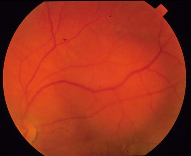
Figure 1: .This is the left fundus of a 55 year old woman with mild blurrying of vision in the right eye for 2 months.
- What is your differential diagnosis of this lesion?
- What ancillary test might be helpful in making the diagnosis?
- What associated finding might you see in the fundus?
- How would you manage this patient?
- Are there any associated systemic findings?
RISPOSTE
Figure 1: .This is the left fundus of a 55 year old woman with mild blurrying of vision in the right eye for 2 months.
- What is your differential diagnosis of this lesion?
Answer: The differential diagnosis of amelanotic lesions of the fundus include choroidal hemangioma, amelanotic melanoma, and metastatic tumors. Given that this is a single, well-circumscribed lesion, metastasis is unlikely. The classic deep orange coloration makes choroidal hemangioma the most likely diagnosis. - What ancillary test might be helpful in making the diagnosis?
Answer: A-scan ultrasonography is useful in differentiating between circumscribed hemangioma and amelanotic melanoma. Although there are variations, choroidal hemangioma often shows high reflectivity whereas amelanotic melanoma typically shows low reflectivity. The A-scan ultrasound in this case was consistent with choroidal hemangioma. - What associated finding might you see in the fundus?
Answer: There may be serous retinal detachment overlying or adjacent to the choroidal hemangioma. - How would you manage this patient?
Answer: If the patient is asymptomatic, no treatment is necessary. If the associated retinal detachment causes visual loss secondary to foveal involvement, photocoagulation is considered with argon laser. The intent is to create chorioretinal adhesion and resolution of the subretinal fluid. - Are there any associated systemic findings?
Answer: Unlike diffuse choroidal hemangioma, which is often associated with nevus flammeus, congenital glaucoma, and ipsilateral intracranial hemangioma, circumscribed choroidal hemangioma is not associated with any systemic findings.