DOMANDA
n. 7
Scott Burk, MD/PhD
Massachusetts Eye and Ear Infirmary, Harvard Medical School, Boston, MA
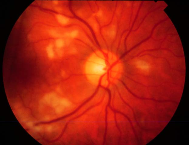
Fig. 1

Fig. 2
Figure 1 and 2: Fundus photographs OD and OS of a 39 year old alcoholic woman admitted to the hospital with abdominal pain. She complains of blind spots near her central vision.
- Given the history, what is the diagnosis?
- What are other causes of this condition?
- What are the fluorescein findings in this condition?
- What is the pathophysiology of this condition?
- What is the treatment for this condition?
RISPOSTE
- Given the history what is the diagnosis?
Answer: This patient was admitted with acute pancreatitis and developed multiple cotton wool spots localized primarily to the posterior pole of both eyes. This represents a non-traumatic presentation of Purtscher's retinopathy - What are other causes of this condition?
Answer: Otmar Purtscher in 1910 described a traumatic retinal angiopathy in patients who had sustained head trauma. The funduscopic findings of multiple cotton wool spots in the posterior pole with or without associated intraretinal hemorrhage has been described in many other conditions including, compressive chest trauma, long bone fracture with fat embolism, amniotic fluid embolism, and in connective tissue disorders such as systemic lupus erythematosis, and scleroderma. - What are the fluorescein findings in this condition?
Answer: The fluorescein angiogram would likely show areas of capillary non-perfusion as well as vessel staining and leakage. - What is the pathophysiology of this condition?
Answer: Purtscher's retinopathy is a retinal vasso-occlusive angiopathy, the exact mechanism of which is not clear. Some theories suggest that fat, air, or even leukocyte emboli result in the blockage of the preterminal retinal arterioles. It is known that complement activation and complement induced leukocyte aggregation occur conditions as diverse as trauma, pancreatitis, and connective tissue diseases. - What is the treatment for this condition?
Answer: The treatment is observation and supportive care. Visual outcome depends upon the severity and location of the nerve fiber infarcts.